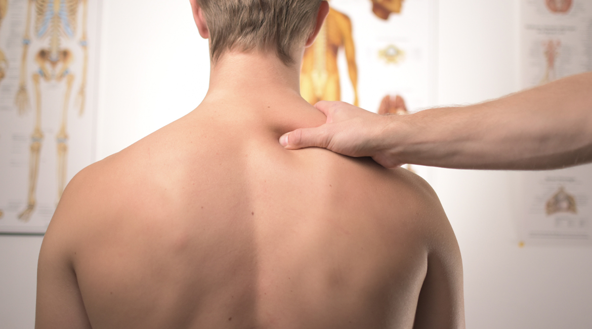
Nowadays the so-called "BACK PAIN" represents a real social plague. It is no coincidence that it represents the first cause of absenteeism at work and it is estimated that around 70-90% of people will have at least one episode of back pain during their lifetime. Before talking about the pathological aspects it is good to give some simple notions of anatomy and physiology.
The vertebral column is formed by 33-34 bone segments: the vertebrae. Each vertebra consists of an anterior portion, the body, and a posterior portion, the arch. The two portions are joined by the peduncles.
As a whole the column is divided into several sections: • Cervical: it includes the first seven vertebrae (C1 to C7). The bone segments of this section have a smaller body than the other portions; furthermore the first two vertebrae have a particular anatomical structure. The first vertebra, called Atlas like the Titan who held the Earth on his shoulders, is articulated with the skull.
• Thoracic: it is formed by 12 vertebrae (T1 to T12). The first 10 vertebrae articulate with the ribs, which form the thoracic cage combined with the sternum. The last 2 vertebrae are articulated with floating ribs that do not articulate with the sternum.
• Lumbar: it consists of 5 vertebrae (L1 to L5). They have a larger body than the aforementioned vertebrae.
• Sacral: the sacrum. It consists of several vertebrae fused together, creating a triangular shape with a downward apex.
• Coccyx: it is a triangular-shaped small bone with a downward apex, given by the fusion of 4-5 vertebrae.
As a whole, the vertebral column is the bearing axis of the human body and responds to two mechanical requirements, rigidity and elasticity, through its 4 curves:
1. The cervical curve with anterior convexity.
2. The dorsal curve with posterior convexity.
3. The lumbar curve with anterior convexity.
4. The sacral curve at the posterior convexity.
The curves increase tenfold the load resistance of the vertebral column. The vertebrae are tied together by joints. The bodies articulate with each other through symphyses, including a fibro-cartilaginous disk (intervertebral disk) consisting of 2 parts:
• annulus fibrosus (external);
• nucleus pulposus (internal): made of 90% of water.
Posteriorly the upper and lower vertebrae articulate with each other by plain joints, which allow the sliding and gliding movements. The anterior longitudinal ligament extends from the skull to the sacrum in front of the vertebral bodies. The posterior longitudinal ligament extends to the vertebral bodies, within the vertebral canal. Stability and mobility are ensured by the proper muscles of the column which are arranged in 2 layers: superficial and deep. The greatest muscular protection is found at the lumbar level.
• The overlap of the vertebrae determines the vertebral canal, where the spinal cord is located. The spinal cord provides the nerves that allow movements and nerves that carry sensory information to the brain. The spinal cord, or spinal marrow, is covered by the meninges and extends into the adult down to L1-L2, after which the cauda equina originates. The cauda equina (horse’s tail) is a bundle of nerves and not a real structure of the nervous system.
The column has three functions: static, dynamic and neuroprotective.
1. Static: it allows us to stand upright in rest conditions.
2. Dynamic: it allows movement through vertebrae and ligaments, the passive mechanical limitation to the amplitude of segmental excursions, and through muscles, which represent the active component.
3. Neuroprotective: it protects the spinal cord.
The column according to the different segments has certain movement abilities or degrees of freedom that cannot be overcome, damage would incur otherwise. • C0-C1-C2: rotation of the head.
• C3 to C7: flexion and extension of the upper limbs. They are the most mobile vertebrae.
• T1 to T10: the vertebrae are immobile due to the joints with the thoracic cage.
• T11-T12: articulated with floating ribs.
• L1 to L5: mobile vertebrae.
• Sacral and coccygeal vertebrae: they are immobile because they are fused. The cervical vertebrae are much smaller and thinner than the lumbar vertebrae, so in the event of an accident they are the most affected. Furthermore, being in a passage area between a mobile and an immobile area, they will be the point of greatest application of the kinetic energy developed in the impact. As a result the damage at this level will be greater. It is also known that the higher the spinal cord injury is, the more severe the damage will be. A cervical damage causes a tetraplegia, a lumbar damage causes paraplegia. A separate chapter in the etiopathogenesis of back pain is dedicated to degenerative diseases.
By definition, a degenerative disease of the spine is an alteration of the vertebrae, discs and ligaments due to their aging processes. As for traumas, the most affected segments are the mobile ones; however these diseases develop independently of muscle protection. This explains why the most involved vertebrae are those in the lumbar section, even though they are the most reinforced from a muscular point of view.
The most frequent pathologies are:
1. Discopathy
2. Disc herniation
3. Spondylosis
The predisposing factors can be congenital (Marfan's syndrome, Wilson's disease ...) or acquired (constitutional, professional, morbid like gout or hypo-hyperparathyroidism).
1. Discopathy: it is a disorder mostly determined by an impoverishment of the watery component of the disc. A herniated disc may develop. It is easier for a hernia to appear directly in young patients. The most affected section is the lumbar one, but this does not exclude the possibility of a discopathy at a higher level. The pivotal symptomatology is given by low back pain that is the most frequent back pain, not associated with pain radiated to one of the legs. This symptomatology regresses with rest. Current medicine proposes: rest, the use of drugs, postural rehabilitation and if strictly necessary a surgical solution that involves the replacement of the disc with a prosthesis. The prostheses, however, stiffen the spine, causing a stressful situation in the vertebrae. A constant and focused yogic practice solves the problem and prevents the aggravation which can lead to a veritable vertebral instability.
2. Herniated disc: certainly the most known among these diseases. It can occur in every section of the spine, but if it occurs at the lumbar level, in addition to low back pain, it can cause what doctors call "radicular pain". This pain tends to radiate to specific regions of one of the legs depending on the compressed nerve root.
The lumbar tracts most affected by the hernia are: L3-L4, L4-L5, L5-S1.
The predisposing factors include:
• Those related to the constitution, such as obesity.
• Those related to the profession: unskilled workers, bellhops, heavy vehicle drivers who often report pain when they take their foot off the clutch.
• Those caused by a decrease in the blood supply to the disc (e.g. due to atherosclerotic processes in the blood vessels), which becomes less rich in water and less elastic. This determines the formation of spaces at the level of the annulus fibrosus, which can come together towards the periphery and determine the expulsion of the nucleus pulp. • Familiarity.
• Postural imbalances.
When a herniated disc develops, the nucleus pulposus of the intervertebral disc herniates posteriorly and protrudes into the vertebral canal. This happens because the annulus fibrosus no longer holds the nucleus pulposus, determining its herniation. The hernia can strike against the roots of a nerve inside the dural sac determining:
• lumbocruralgia, lumbar pain radiated to the groin and down to the medial part of the knee if the herniated disc is L3-L4;
• lumbosciatalgia, pain that descends posteriorly and laterally or only posteriorly to the foot if the herniated disc is L4-L5 or L5-S1.
Generally, the onset of a hernia is sudden, accompanied by acute, well-localized pain. In the following hours the pain increases until it becomes continuous. This occurs because the ligaments and tissues damaged by the protrusion become congested with blood, attracting inflammatory mediators. In addition, the muscles of the affected area contract to prevent painful movements. Very often the hernia can regress, even spontaneously disappear, since the herniated fragment undergoes a drying process or phagocytosis, i.e. elimination by the immune system. Conservative and surgical therapy are available. The first is used in the initial phase of the disease: an elastic band is used in order to alleviate pain, but it has no healing prospects; anti-inflammatories and analgesics are administered, a physio-kinesis therapy is proposed. Surgical treatment is only used when severe neurological deficits are present (e.g. cauda equina syndrome or major strength defects in the lower limbs) or when conservative treatment continued for at least 4-6 weeks has not alleviated the symptoms. The practice of yoga facilitates and accelerates the healing process by avoiding surgery and its complications; obviously the guidance of a competent instructor is required. Relapse, frequent in those suffering from these pathologies, is prevented with a regular and growing practice.
3. Spondylosis, or spondylodiscoarthrosis: less frequent than the herniated disc, it mainly affects subjects between 50 and 70 years old. To better understand what we are talking about we must define the concept of arthrosis: "It is a chronic evolutionary arthropathy that involves a regression of the structures that make up the joint. The production of osteophytes is a characteristic element: bones are rearranged at the edges of the joints." The spondylodiscoarthrosis is a degenerative process of the bone component and discal spine with the production of osteophytes that determine a narrowing (stenosis) of the vertebral canal. It can strike at any level of the column. If the lumbar portion is affected, the lumbar pain will be radiated to the buttocks and legs. The characteristic symptom is the neurogenic claudication which consists in the presence of pain and tingling in the back and legs which tends to decrease with rest. If the osteophytes compress the nerve roots, there will be a symptomatology similar to that of the herniated disc with lumbocruralgia or lumbosciatalgia depending on the segment involved.
In any case, the practice of yoga is particularly useful, both in terms of prevention and from a therapeutic point of view. Yoga regenerates the damaged tissues restoring the normal physiology of the spine, if taught according to the original rules as transmitted by the ancient texts and masters. Otherwise it becomes a discipline that can cause serious damage, instead of improving our health. For this reason it is always good to rely on someone competent rather than on the first yoga course you might find.
The vertebral column is formed by 33-34 bone segments: the vertebrae. Each vertebra consists of an anterior portion, the body, and a posterior portion, the arch. The two portions are joined by the peduncles.
As a whole the column is divided into several sections: • Cervical: it includes the first seven vertebrae (C1 to C7). The bone segments of this section have a smaller body than the other portions; furthermore the first two vertebrae have a particular anatomical structure. The first vertebra, called Atlas like the Titan who held the Earth on his shoulders, is articulated with the skull.
• Thoracic: it is formed by 12 vertebrae (T1 to T12). The first 10 vertebrae articulate with the ribs, which form the thoracic cage combined with the sternum. The last 2 vertebrae are articulated with floating ribs that do not articulate with the sternum.
• Lumbar: it consists of 5 vertebrae (L1 to L5). They have a larger body than the aforementioned vertebrae.
• Sacral: the sacrum. It consists of several vertebrae fused together, creating a triangular shape with a downward apex.
• Coccyx: it is a triangular-shaped small bone with a downward apex, given by the fusion of 4-5 vertebrae.
As a whole, the vertebral column is the bearing axis of the human body and responds to two mechanical requirements, rigidity and elasticity, through its 4 curves:
1. The cervical curve with anterior convexity.
2. The dorsal curve with posterior convexity.
3. The lumbar curve with anterior convexity.
4. The sacral curve at the posterior convexity.
The curves increase tenfold the load resistance of the vertebral column. The vertebrae are tied together by joints. The bodies articulate with each other through symphyses, including a fibro-cartilaginous disk (intervertebral disk) consisting of 2 parts:
• annulus fibrosus (external);
• nucleus pulposus (internal): made of 90% of water.
Posteriorly the upper and lower vertebrae articulate with each other by plain joints, which allow the sliding and gliding movements. The anterior longitudinal ligament extends from the skull to the sacrum in front of the vertebral bodies. The posterior longitudinal ligament extends to the vertebral bodies, within the vertebral canal. Stability and mobility are ensured by the proper muscles of the column which are arranged in 2 layers: superficial and deep. The greatest muscular protection is found at the lumbar level.
• The overlap of the vertebrae determines the vertebral canal, where the spinal cord is located. The spinal cord provides the nerves that allow movements and nerves that carry sensory information to the brain. The spinal cord, or spinal marrow, is covered by the meninges and extends into the adult down to L1-L2, after which the cauda equina originates. The cauda equina (horse’s tail) is a bundle of nerves and not a real structure of the nervous system.
The column has three functions: static, dynamic and neuroprotective.
1. Static: it allows us to stand upright in rest conditions.
2. Dynamic: it allows movement through vertebrae and ligaments, the passive mechanical limitation to the amplitude of segmental excursions, and through muscles, which represent the active component.
3. Neuroprotective: it protects the spinal cord.
The column according to the different segments has certain movement abilities or degrees of freedom that cannot be overcome, damage would incur otherwise. • C0-C1-C2: rotation of the head.
• C3 to C7: flexion and extension of the upper limbs. They are the most mobile vertebrae.
• T1 to T10: the vertebrae are immobile due to the joints with the thoracic cage.
• T11-T12: articulated with floating ribs.
• L1 to L5: mobile vertebrae.
• Sacral and coccygeal vertebrae: they are immobile because they are fused. The cervical vertebrae are much smaller and thinner than the lumbar vertebrae, so in the event of an accident they are the most affected. Furthermore, being in a passage area between a mobile and an immobile area, they will be the point of greatest application of the kinetic energy developed in the impact. As a result the damage at this level will be greater. It is also known that the higher the spinal cord injury is, the more severe the damage will be. A cervical damage causes a tetraplegia, a lumbar damage causes paraplegia. A separate chapter in the etiopathogenesis of back pain is dedicated to degenerative diseases.
By definition, a degenerative disease of the spine is an alteration of the vertebrae, discs and ligaments due to their aging processes. As for traumas, the most affected segments are the mobile ones; however these diseases develop independently of muscle protection. This explains why the most involved vertebrae are those in the lumbar section, even though they are the most reinforced from a muscular point of view.
The most frequent pathologies are:
1. Discopathy
2. Disc herniation
3. Spondylosis
The predisposing factors can be congenital (Marfan's syndrome, Wilson's disease ...) or acquired (constitutional, professional, morbid like gout or hypo-hyperparathyroidism).
1. Discopathy: it is a disorder mostly determined by an impoverishment of the watery component of the disc. A herniated disc may develop. It is easier for a hernia to appear directly in young patients. The most affected section is the lumbar one, but this does not exclude the possibility of a discopathy at a higher level. The pivotal symptomatology is given by low back pain that is the most frequent back pain, not associated with pain radiated to one of the legs. This symptomatology regresses with rest. Current medicine proposes: rest, the use of drugs, postural rehabilitation and if strictly necessary a surgical solution that involves the replacement of the disc with a prosthesis. The prostheses, however, stiffen the spine, causing a stressful situation in the vertebrae. A constant and focused yogic practice solves the problem and prevents the aggravation which can lead to a veritable vertebral instability.
2. Herniated disc: certainly the most known among these diseases. It can occur in every section of the spine, but if it occurs at the lumbar level, in addition to low back pain, it can cause what doctors call "radicular pain". This pain tends to radiate to specific regions of one of the legs depending on the compressed nerve root.
The lumbar tracts most affected by the hernia are: L3-L4, L4-L5, L5-S1.
The predisposing factors include:
• Those related to the constitution, such as obesity.
• Those related to the profession: unskilled workers, bellhops, heavy vehicle drivers who often report pain when they take their foot off the clutch.
• Those caused by a decrease in the blood supply to the disc (e.g. due to atherosclerotic processes in the blood vessels), which becomes less rich in water and less elastic. This determines the formation of spaces at the level of the annulus fibrosus, which can come together towards the periphery and determine the expulsion of the nucleus pulp. • Familiarity.
• Postural imbalances.
When a herniated disc develops, the nucleus pulposus of the intervertebral disc herniates posteriorly and protrudes into the vertebral canal. This happens because the annulus fibrosus no longer holds the nucleus pulposus, determining its herniation. The hernia can strike against the roots of a nerve inside the dural sac determining:
• lumbocruralgia, lumbar pain radiated to the groin and down to the medial part of the knee if the herniated disc is L3-L4;
• lumbosciatalgia, pain that descends posteriorly and laterally or only posteriorly to the foot if the herniated disc is L4-L5 or L5-S1.
Generally, the onset of a hernia is sudden, accompanied by acute, well-localized pain. In the following hours the pain increases until it becomes continuous. This occurs because the ligaments and tissues damaged by the protrusion become congested with blood, attracting inflammatory mediators. In addition, the muscles of the affected area contract to prevent painful movements. Very often the hernia can regress, even spontaneously disappear, since the herniated fragment undergoes a drying process or phagocytosis, i.e. elimination by the immune system. Conservative and surgical therapy are available. The first is used in the initial phase of the disease: an elastic band is used in order to alleviate pain, but it has no healing prospects; anti-inflammatories and analgesics are administered, a physio-kinesis therapy is proposed. Surgical treatment is only used when severe neurological deficits are present (e.g. cauda equina syndrome or major strength defects in the lower limbs) or when conservative treatment continued for at least 4-6 weeks has not alleviated the symptoms. The practice of yoga facilitates and accelerates the healing process by avoiding surgery and its complications; obviously the guidance of a competent instructor is required. Relapse, frequent in those suffering from these pathologies, is prevented with a regular and growing practice.
3. Spondylosis, or spondylodiscoarthrosis: less frequent than the herniated disc, it mainly affects subjects between 50 and 70 years old. To better understand what we are talking about we must define the concept of arthrosis: "It is a chronic evolutionary arthropathy that involves a regression of the structures that make up the joint. The production of osteophytes is a characteristic element: bones are rearranged at the edges of the joints." The spondylodiscoarthrosis is a degenerative process of the bone component and discal spine with the production of osteophytes that determine a narrowing (stenosis) of the vertebral canal. It can strike at any level of the column. If the lumbar portion is affected, the lumbar pain will be radiated to the buttocks and legs. The characteristic symptom is the neurogenic claudication which consists in the presence of pain and tingling in the back and legs which tends to decrease with rest. If the osteophytes compress the nerve roots, there will be a symptomatology similar to that of the herniated disc with lumbocruralgia or lumbosciatalgia depending on the segment involved.
In any case, the practice of yoga is particularly useful, both in terms of prevention and from a therapeutic point of view. Yoga regenerates the damaged tissues restoring the normal physiology of the spine, if taught according to the original rules as transmitted by the ancient texts and masters. Otherwise it becomes a discipline that can cause serious damage, instead of improving our health. For this reason it is always good to rely on someone competent rather than on the first yoga course you might find.